
SHIN LABORATORY
The Shin Lab uses a multi-disciplinary approach to develop nano-biomaterials combined with micro and nano fabrication technologies for engineering tissue or organs for several biomedical applications.
Electrically Conductive Materials for Cardiac Tissue Engineering
 Cardiac tissue engineering aims at assembling tissue constructs that can restore basic cardiac function by incorporating cellular components within scaffolds. To address these challenges, extracellular matrix (ECM) based biomaterials have been widely used to incorporate cardiovascular cells, since the hydrogels closely mimic the natural ECM and can be designed to present soluble and insoluble factors to optimize cellular behaviors. However, a key limitation of current ECM-based biomaterials is limited cell-cell coupling and delayed electrical signal propagation due to the lack of their electrical conductivity. Electrically conductive nanoparticles with unique physical and chemical properties can be incorporated to create electrically active ECM which leading to improved cell adhesion, organization, and cell-cell coupling. The electrically conductive networks within the implant will allow for adequate physiological electrical propagation to pass through the implant area in large-scale cardiac injury.
Development of Advanced Microfabrication Techniques
 Our lab uses Rousselot low endotoxin GelMA gelatins in 3D bioink formulations as cell supporting matrix for tissue engineering.
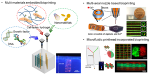
Recent advances in microfabrication techniques have enabled researchers to build increasingly complex architectures with tunable biophysical properties. These technologies offer significant potential in tissue engineering and regenerative medicine and enable us to develop highly-organized and functional three-dimensional (3D) constructs that mimic the architectural complexity of in vivo organ systems. Specially, bioprinting has emerged as a technology that can fabricate biomimicked 3D tissue constructs. Consequently, our team has employed, adapted, and invented a wide range of 3D bioprinting strategies integrated with a microfluidic printhead, multi-axial nozzle, or self-healing hydrogel bath to build tissue scaffolds capable of recapitulating key features of in vivo tissue and organ physiology.
Manufacturing Biomimetic Scaffolds to Promote Skeletal Muscle Regeneration (Stepping Strong Innovator Project)
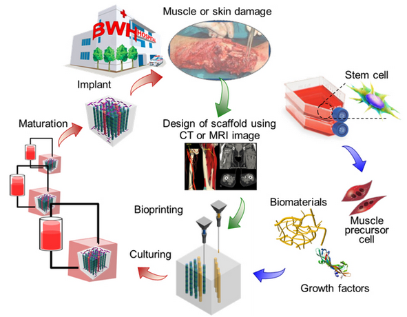 Approximately 500,000 patients in the US suffer from volumetric muscle loss (VML) injuries every year. VML usually results from traumatic events, such as motor-vehicle accidents or bomb blasts, in which a significant portion of a muscle or group of muscles are lost or irreversibly damaged. Following this, the injured area heals by substantial scarring rather than the regeneration of skeletal muscle, with a subsequent loss of muscle strength and function. Patients affected by VML injuries return to work only in ~50% of cases. VML injuries can overwhelm the normally robust regenerative capacity of skeletal muscle. Therefore, these injuries heal with minimal muscle regeneration and substantial fibrosis, thereby leading to impaired muscle function. Our strategy is to thereby create a customized scaffold which (1) mimics muscle architecture and mechanical properties of patient muscle tissue, (2) incorporates growth factors and (3) deliver muscle stem cells to traumatic muscular injuries following volumetric muscle loss.
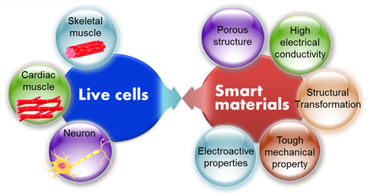
Bio-hybrid Actuators
Engineered living-synthetic systems with a capacity to dynamically deform their shape and sense biological environments have been developed for various biomedical applications, such as building biorobots, biosensors, and artificial muscles. Despite the significant advances in this field, many limitations still remain, such as creating life-like movements, on-board control systems, autonomous control over direction and speed, robust actuation, and low-power stimulation systems for the biorobots. To overcome these limitations, living muscle actuator technologies have borrowed inspiration from biomimetic concepts to develop bio-inspired robots. Our research team is interested in developing a bio-inspired soft robotics system, with integrated self-actuating cardiac muscles on a hierarchically structured scaffold with flexible microelectrodes.
Organ-on-a-chip Systems
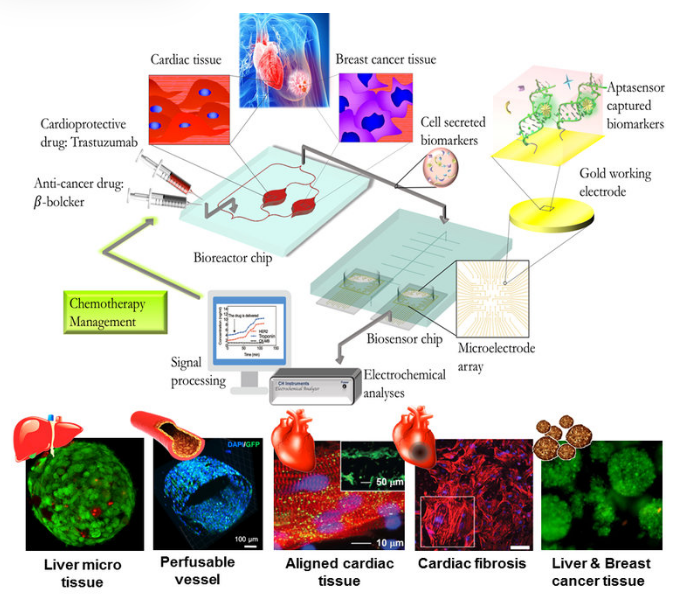 Organ-on-a-chip systems are miniaturized microfluidic 3D human tissue and organ models designed to recapitulate the important biological and physiological parameters of their in vivo counterparts. They have recently emerged as a viable platform for personalized medicine and drug screening. Our research team has been developing organ-on-a-chip platform, a modular, scalable microfluidic system, and biosensor with multi-tissue system integration (mimicking human organ function), which can be achieved by the creation of resealable microfluidic devices capable of cell culture and analysis of cell secreted biomarkers.
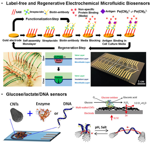
Biosensors and Flexible Bioelectronics
 Organ-on-a-chip systems are miniaturized microfluidic 3D human tissue and organ models designed to recapitulate the important biological and physiological parameters of their in vivo counterparts. They have recently emerged as a viable platform for personalized medicine and drug screening. Our research team has been developing organ-on-a-chip platform, a modular, scalable microfluidic system, and biosensor with multi-tissue system integration (mimicking human organ function), which can be achieved by the creation of resealable microfluidic devices capable of cell culture and analysis of cell secreted biomarkers.